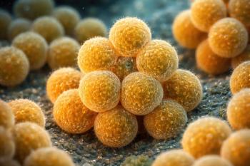
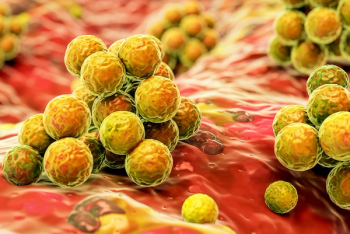
Rifampin (or Rifampicin) is a class of antibiotics that is mostly associated as part of the 4-drug regimen for active tuberculosis (TB) and as monotherapy for latent TB.1,2 Outside of TB, it’s been traditionally used for bone, joint, and hardware infections with other antibiotics and not as monotherapy due to the rapid emergence of resistance via a single mutation in the rpoB gene.3 Rifampin works by inhibiting DNA-dependent RNA polymerase preventing bacterial RNA synthesis.2 It is particularly bactericidal against Staphylococcal aureus infections due to its ability to achieve high intracellular levels, kill organisms in the sessile phase of growth (non-replicating bacteria) and penetrate biofilms.4 Its “anti-biofilm” properties have been closely linked to its use for years. But what exactly is a biofilm? How is it related to S aureus infections? What is its role in bone, joint, and hardware infections?
The “Infamous” Biofilm
The first description of biofilms dates as far back as the 17th century "the Father of Microbiology", Anton Von Leeuwenhoek, observed microbial aggregates (“animalcules”) on the plaque taken from teeth.5 At that time, it was not called biofilm yet, but it laid the foundation of how we describe biofilms today. The term itself was first coined in 1978 by J. William Costerton in a paper titled “How Bacteria Stick”- a simple but fitting description of how a “mass of tangled fibers of polysaccharides, or branching sugar molecules, that extend from the bacterial surface and form a feltlike ‘glycocalyx’ surrounding an individual cell or a colony of cells” help bacteria survive, propagate and resist antibiotics6. Donlan and Costerton in a 2002 paper2 described biofilm as a “microbially derived sessile community characterized by cells that are irreversibly attached to a substratum or interface or to each other, are embedded in a matrix of extracellular polymeric substances that they have produced and exhibit an altered phenotype with respect to growth rate and gene transcription.”7 This partly explained biofilm’s resistance against antibiotics. Biofilm-associated cells grow slower than planktonic cells and, as a result, take up antibiotics more slowly.7 This acts as diffusion barriers reducing antimicrobial penetrance towards the deeper layers of the biofilm.8 This makes bacteria within biofilms 10 to 1000 times more resistant compared to bacteria not associated with biofilms. Altered metabolic activity of biofilm bacteria is also hypothesized to be a major driver of resistance in addition to decrease in antimicrobial penetration. These 2 mechanisms contribute to the increased risk of persistence and recurrence of infections, specifically those caused by S aureus.
Osteomyelitis (OM)
The use of rifampin had been explored as an adjunctive therapy for OM even in the absence of any prosthesis or hardware to reduce the risk of recurrence and treatment failure.8,9,10 It has excellent bone penetration reaching mean bone concentrations 1.3±1 µg/mL and 6.5±1.3 µg/mL in cortical and cancellous bones, respectively.11 This would reach levels higher than the minimum inhibitory concentration susceptibility breakpoint of rifampin against S aureus of ≤1 µg/mL.
Biofilms that are historically formed on infected hardware are also observed in bone infections, specifically chronic OM. The reduction in the penetration of antibiotics encourages the formation of small colony variants (SCVs) and persisters.8 SCVs are small sized colonies characterized by slow growth, downregulation of virulence factors and upregulation of genes associated with adhesion and biofilm formation. Persisters are dormant variants found within a susceptible bacterial population with increased antibiotic tolerance. These two phenomena are indicative of the changes in the metabolic activity of bacteria within biofilms causing increased antibiotic tolerance. These support the enduring and relapsing nature of chronic OM.
What You Need to Know
Biofilms are complex microbial communities embedded in a matrix of extracellular substances, contributing to bacterial resistance against antibiotics. Rifampin's ability to penetrate biofilms makes it effective against Staphylococcal aureus infections, particularly in bone, joint, and hardware infections.
Rifampin plays a significant role in adjunctive therapy for osteomyelitis and prosthetic joint infections (PJIs). Studies suggest potential benefits, such as lower rates of amputation and death in diabetic foot osteomyelitis patients receiving rifampin.
While rifampin has demonstrated efficacy in certain infections, its use requires careful consideration of factors such as dosing, timing, and potential drug interactions.
In 2019, Wilson et al conducted an observational cohort study using the Veterans Health Administration database to evaluate the role of adjunctive rifampin in preventing subsequent amputations and deaths in patients with diabetic foot OM (DFO).10 Lower rates of amputation and death were observed among patients receiving rifampin (35 of 130 [26.9%] vs. 2250 of 6044 [37.2%], P=0.02). Several statistically significant differences between the groups may have affected the outcome given that patients who were given rifampin were younger, had fewer comorbidities, more infectious disease specialist involvement, and more S aureus identified on cultures. A meta-analysis of 4 randomized controlled trials (RCT) (Van der Auwera et al12, Norden et al13, Zimmerli et al14, and Karlsen et al15) suggested a potential benefit of adjunctive rifampin in patients with OM with or without hardware.9 Van der Auwera et al and Norden et al did not include any hardware while Zimmerli et al and Karlsen et al were studies involving prosthetic joints. It is important to point out the included studies had small sample sizes, different dosing regimens for rifampin, differences in coantibiotics utilized, heterogeneity of patient population, and differences in surgical management.
While the potential advantages might be more noticeable in individuals with chronic OM and DFO, a clear recommendation regarding its use cannot be established in the absence of well-designed RCTs specifically addressing this issue.
Prosthetic Joint Infections (PJIs)
Staphylococcal PJIs have a heterogenous success rate ranging from 23% to 90%.16 This variability in success rates leads to differing opinions on PJI treatment specifically when it comes to antibiotic management. One of the long standing tenets of PJI, and hardware infections in general, is the addition of rifampin to standard antibiotic management. PJIs occur in 1%-2% of all patients following a large joint replacement.17 This carries with it considerable comorbidities, decreased quality of life, as well as increased costs both to the patient and the healthcare system in general.1,17
Debridement, antibiotics, and implant retention (DAIR) has been the surgical intervention of choice for acute PJIs. The variability in the definition of what “acute” means ranges from 30 to 90 days after index arthroplasty, or with an acute hematogenous PJI with symptoms for < 3 weeks.15,17,18 Despite this, DAIR failure rates are reported to be ranging from 10% to 45%16. The Infectious Diseases Society of America published in 2012 the clinical practice guidelines on PJI and recommended rifampin for susceptible Staphylococcal PJI following debridement and retention of prosthesis.1 This was influenced by the landmark RCT by Zimmerli et al (1998) showing combination therapy with ciprofloxacin and rifampin resulted in similar cure rates compared to ciprofloxacin monotherapy.14 The study specifically included only those with retained stable prosthesis. Unfortunately, the sample size was small (33 total patients, 24 of whom fully completed the trial) and the use of ciprofloxacin monotherapy as the comparator had been called into question due to 4/5 failures were due to ciprofloxacin resistance.
The role of rifampin in Staphylococcal PJI has been called into question by a RCT conducted by Karlsen et al that showed no significant difference in remission rate between the rifampin combination group (74%, 17/23) and the monotherapy group (72%, 18/25).14 Unfortunately, the study was underpowered to draw any definitive conclusion, albeit having a larger number of patients included in the final analysis (48 patients in Karlsen et al15 vs 24 patients in Zimmerli et al14). So far, these are the only 2 RCTs looking at the role of rifampin for PJIs while the rest are observational studies.16,19
One of those observational studies was conducted Davis et al in 27 hospitals across Australia and New Zealand that aimed to look at predictors of treatment success after PJI.17 DAIR was the main surgical treatment in 352/653 patients where 51% used rifampin (61% where ≥1 causative organisms were gram-positive coccus, and 75% where ≥1 organism was Staphylococcus). The use of rifampin was not independently associated with treatment success (OR = 1.15; 95% CI, 0.82–1.61). These findings were consistent if the analysis is restricted to gram-positive infections, Staphylococcal infections, or those with Staphylococcal infections who received ≥14 days of rifampin. Younger age, hip as the index joint, early infection (<30 days from original arthroplasty), shorter duration of symptoms at diagnosis, high baseline albumin, and absence of chronic renal disease or malignancy were independently associated with treatment success. While knee as the index joint, certain organisms (S aureus, Cutibacterium spp., or gram-negative rods) and late infections (>30 days from original arthroplasty) were associated with lower treatment success rates.
There are 2 meta-analyses16,19 that tried to evaluate the utility of rifampin for PJIs. Aydin et al conducted a meta-analysis of 13 observational studies and looked at 3 subsets focusing on Staphylococcal spp. (n=568), Streptococcal spp. (n=483) and Propionibacterium spp. (n=80), which were the predominant bacteria in the studies included19. Only the Streptococcal subset was found to favor the use of rifampin adjunctive regimens. The 1 study included in the meta-analysis that specifically examined groups with or without rifampin found that adjunctive rifampin was not superior. There were several limitations, particularly the quality of evidence ie no RCTs, and all the included studies were compromised by selection bias. The authors concluded that the evidence did not favor the addition of rifampin especially for Staphylococcal and Propionibacterium PJIs.
The 2nd meta-analysis was conducted by Scheper et al and included 64 studies (n=4380) published between 1990 and September 2, 2020.16 There were 62 observational cohort studies (3 prospective, 59 retrospective) and 2 RCTs (Zimmerli et al14 and Karlsen et al).15 Out of the 64 studies, 49 studies use rifampin for Staphylococcus PJI. Thirty of the 49 studies have reported outcomes of treatment with or without rifampin. The pooled success rate in all studies was 60%. If stratified based on the causative organism, the pooled success rates for S aureus PJI and Coagulase-Negative Staphylococcus (CONS) PJI after DAIR were 62% (2922 analyzed patients in 54 studies) and 73% (36 studies, 760 patients), respectively.
The addition of rifampin after DAIR had a higher success rate (64% in 34 studies with 2884 patients) compared to those who were not on rifampin (44% in 18 studies with 976 patients) with similar procedure. Staphylococcal knee PJIs (2 studies) with addition of rifampin had higher cure rates (81%, 56/69) than without rifampin (50% 17/34). This was also observed in 24 studies with Staphylococcal knee and hip PJIs (with rifampin 70%, 903/1298 vs without rifampin 5.3%, 186/354). The 3 studies that specifically looked at combined knee and hip PJIs where S aureus was the causative organism also showed higher cure rates with rifampin (80%, 100/125) than without rifampin (40%, 4/10). Whereas Staphylococcal hip PJIs (4 studies) treated with additional rifampin have similar cure rates compared to those not on rifampin (83%, 102/123 vs. 82%, 28/34).