|Articles|July 13, 2023
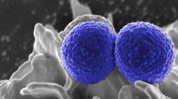
How COVID-19 Infiltrates and Inflames the Heart
Author(s)Nina Cosdon
Neuropilin-1 enables COVID-19 to enter human cardiomyocytes, accounting for increased proteases activity and apoptotic markers that lead to cell damage and apoptosis.
Advertisement
Although primarily associated with respiratory complications, COVID-19 can be severe or fatal for individuals with preexisting heart conditions.
The reasons for COVID-19-related cardiac damage are largely unknown, but inflammation (i.e., cytokine storm) and oxidative stress are believed to be involved.
One
The research team sought to determine if cardiomyocytes were targeted by SARS-CoV-2, and how inflammation and oxidative stress are involved. They compared the impact of pro-inflammatory and oxidative stress on virus entry and virus-associated cardiac damage between COVID-19 patients and non-infected donor hearts.
The investigators found the SARS-CoV-2 virus directly in heart cells. “Our observations show that the virus exerts pressure on the heart muscle, attacks and weakens the contractile force, i.e., the pumping function of the heart,” said Nazha Hamdani, PhD, an author of the study.
The team found the virus may be able to penetrate the heart muscles cells due to increased proteolytic activity, which activates specific enzymes that degrade proteins and lead to cardiac muscle cell dysfunction. The entry of COVID-19 into heart cells results from the activation of the spike protein enzymes responsible for the degradation of proteins, its entry into cells relying on this degradation.
The investigators also examined the proteins responsible for apoptosis, finding that while they had increased activity, their expression was significantly reduced. Hamdani said, “This indicates that the proteins are cleaved, and apoptosis is activated. The results imply that apoptosis contributes to the deterioration in cardiac contractility observed in SARS-CoV-2 patients.”
After exploring what promotes this increased proteolytic activity and apoptosis of cardiac myocytes, the investigators identified COVID-19-associated inflammatory environment and oxidative stress.
Neutrophils, a primary cell type that releases proteolytic enzymes, are rapidly mobilized from the bloodstream into the damaged tissue during an inflammatory response. Proteolytic enzymes are released more frequently in COVID-19 patients, leading the investigators to analyze the signal pathway of the interleukin-6-driven neutrophil traffic.
“We found that neuropilin-1 potentiates SARS-CoV-2 entry into human cardiomyocytes, a phenomenon driven by inflammatory and oxidant signals,” the study authors wrote. “These changes accounted for increased proteases activity and apoptotic markers thus leading to cell damage and apoptosis.”
The team found that inflammatory signaling pathways in cardiac myocytes were highly regulated, and interleukin-6 was highly elevated, suggesting the critical role of white blood cells in COVID-19 patients and their inflamed pathways.
“SARS-CoV-2 is able to spread in the infected heart in a receptor-dependent and receptor-independent manner,” explained Hamdani. “We also examined another mechanism by which the virus can gain access to the heart muscle cells, thus contributing to endothelial dysfunction. We will soon be able to publish these results.”
Advertisement
Related Content
Advertisement


Advertisement
Advertisement
Trending on Contagion Live
1
Important Clinical Practice Takeaways From the DOTs and PeterPen Trials
2
SNAP Global Trial Supports Cefazolin for MSSA Bacteremia
3
10 Infectious Disease Developments That Defined Q2 2026
4
Merck, ADAP Crisis Task Force Agree to Expand IDVYNSO Access in HIV
5