Studies
Laboratory evaluation revealed leukocytosis with neutrophilia (white blood cell count 29.3 K/µL), normocytic anemia (hemoglobin 9.6 g/dL), thrombocytopenia (platelet count 124 × 10³/µL), hypoalbuminemia (albumin 2.3 g/dL), and markedly elevated inflammatory markers, including C-reactive protein (16.0 mg/dL) and procalcitonin (30.1 ng/mL). Viral respiratory panel testing, malaria testing, QuantiFERON-TB, HIV testing, and urinalysis were negative.
A CT of the abdomen and pelvis demonstrated portal vein thrombosis with a small right hepatic lesion, mild splenomegaly, and small-volume ascites. A right upper quadrant ultrasound confirmed portal vein thrombosis. An MRI of the abdomen further characterized several indeterminate right hepatic lobe lesions measuring up to 1.7 cm in the setting of known portal vein thrombus and splenomegaly.
Clinical Course
The patient was admitted, and anticoagulation with intravenous heparin was initiated. Blood cultures obtained during hospitalization revealed polymicrobial bacteremia. Blood cultures collected on hospital day 1 grew Bacteroides fragilis in 2 sets, with 1 set also growing Staphylococcus epidermidis, which was considered a contaminant. Repeat blood cultures obtained on hospital day 3 grew Streptococcus anginosus in 2 sets. Subsequent blood cultures remained negative. Given the bacteremia and concern for infective endocarditis, transthoracic echocardiography was initially performed, demonstrating preserved ejection fraction without obvious valvular vegetations. Transesophageal echocardiography was subsequently performed for further evaluation and revealed a small mobile echodensity measuring approximately 0.5 cm on the posterior leaflet of the mitral valve, with mild mitral regurgitation, raising concern for possible infective endocarditis.
Interventional radiology performed an ultrasound-guided biopsy and aspiration of the hepatic lesion. Aspiration yielded purulent material, and histopathologic evaluation demonstrated hepatocytes in a background of marked acute inflammation without evidence of malignancy. Bacterial cultures from the aspirate were negative. Given the patient’s travel history and imaging findings, parasitic infection was considered. Serologic testing subsequently returned positive for E histolytica IgG antibodies, supporting the diagnosis of invasive amoebiasis with amoebic liver abscess.
The patient was initially treated with broad-spectrum antimicrobial therapy, including meropenem and daptomycin, and was subsequently transitioned to intravenous ceftriaxone and oral metronidazole based on infectious disease recommendations. Given the concern for possible infective endocarditis, a 6-week course of intravenous ceftriaxone 2 g every 24 hours was prescribed, along with a 2-week course of metronidazole. Anticoagulation was transitioned to apixaban for the management of portal vein thrombosis. Cardiothoracic surgery staff evaluated the patient and recommended no surgical intervention. The patient's fevers resolved, leukocytosis normalized, and repeat blood cultures remained negative. He was discharged in stable condition with a peripherally inserted central catheter for outpatient intravenous antibiotic therapy and infectious disease follow-up 1 week after discharge.
Discussion
E histolytica is a protozoan parasite responsible for amoebiasis, an infection that affects approximately 50 million individuals worldwide and causes significant morbidity in endemic regions of Central and South America, Africa, and Asia. Invasive amoebiasis remains an important cause of extraintestinal infection in individuals returning from endemic regions. Following ingestion of cysts in contaminated food or water, E histolytica trophozoites colonize the colon and may invade the intestinal mucosa. In a subset of patients, organisms enter the portal venous circulation and disseminate to the liver, resulting in amoebic liver abscess.1,2 Although intestinal symptoms are common, many patients with extraintestinal disease do not demonstrate clinically apparent colitis at the time of diagnosis, which can complicate recognition of invasive infection.3
Amoebic liver abscess is the most common extraintestinal manifestation of amoebiasis and typically presents with fever and systemic inflammatory symptoms. Imaging often demonstrates solitary or multiple lesions in the right hepatic lobe.2 Aspiration may yield purulent material; however, bacterial cultures frequently remain negative because the underlying infection is protozoal rather than bacterial. As a result, serologic testing plays an important diagnostic role, particularly in patients with compatible travel history and imaging findings.1,3 In the present case, the patient’s positive E histolytica IgG serology, together with recent travel to Peru and exposure to potentially contaminated freshwater in the Amazon basin, supported the diagnosis of invasive amoebiasis with hepatic abscess formation.
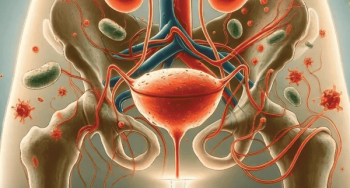
Pylephlebitis, or septic thrombophlebitis of the portal venous system, is a rare but potentially life-threatening complication of intra-abdominal infection. It occurs when infection spreads into the mesenteric venous circulation and propagates to the portal vein, resulting in thrombosis and bacteremia.4, 6 Historically, pylephlebitis has been associated with appendicitis and diverticulitis; however, hepatic abscess and other intra-abdominal infections have also been implicated.4 Portal vein thrombosis in this patient likely developed in association with hepatic abscess formation and portal venous infection.
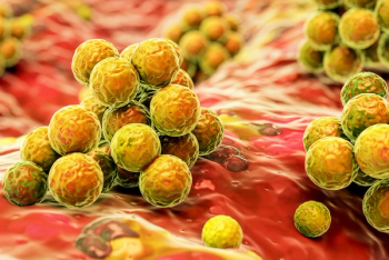
The patient subsequently developed polymicrobial bacteremia involving B fragilis and S anginosus, organisms commonly associated with gastrointestinal infections and intra-abdominal abscesses. Persistent bacteremia prompted evaluation for infective endocarditis, and transesophageal echocardiography demonstrated a small mobile echodensity on the posterior leaflet of the mitral valve, concerning for a possible vegetation. Infective endocarditis can develop when circulating pathogens adhere to endocardial surfaces and form vegetations.5 There are no reported cases of E histolytica infective endocarditis; therefore, the patient's valvular findings were more likely attributable to the documented bacterial bloodstream infection than to direct amoebic involvement.
An important diagnostic consideration in this case is the distinction between an amoebic liver abscess with secondary bacterial translocation and a primary pyogenic liver abscess with incidental positive amoebic serology. Although E histolytica IgG serology positivity supports prior or current exposure, it does not definitively establish the organism as the direct cause of the hepatic abscesses. Pyogenic liver abscesses associated with organisms such as S anginosus and B fragilis may originate from gastrointestinal sources and present with similar clinical and radiographic findings. Amoebic liver abscesses typically yield sterile bacterial cultures unless secondary bacterial infection is present, whereas pyogenic abscesses more commonly demonstrate bacterial growth.2 Given the positive amoebic serology and concurrent polymicrobial bacteremia, an amoebic liver abscess complicated by secondary bacterial infection remains a plausible explanation, although a primary pyogenic process cannot be definitively excluded.
The sequence observed in this case illustrates a potential portal–systemic infectious cascade, in which invasive intestinal infection results in hepatic abscess formation through portal venous dissemination. Local infection within the portal circulation may then lead to septic portal vein thrombosis, followed by systemic bacteremia and distant complications such as infective endocarditis. While individual components of this sequence have been described in the literature, reports describing the coexistence of invasive amoebiasis, pylephlebitis, polymicrobial bacteremia, and cardiac involvement remain exceedingly uncommon.4,7-9 Recognition of this potential progression is important in patients presenting with hepatic lesions and portal vein thrombosis in the context of relevant travel exposure, as early diagnosis and treatment may prevent systemic complications.
References
1. Shirley DAT, Farr L, Watanabe K, Moonah S. A review of the global burden, new diagnostics, and current therapeutics for amebiasis. Open Forum Infect Dis. 2018;5(7):ofy161. doi:10.1093/ofid/ofy161
2. Stanley SL Jr. Amoebiasis. Lancet. 2003;361(9362):1025-1034. doi:10.1016/S0140-6736(03)12830-9
3. Haque R, Huston CD, Hughes M, Houpt E, Petri WA Jr. Amebiasis. N Engl J Med. 2003;348(16):1565-1573. doi:10.1056/NEJMra022710
4. Plemmons RM, Dooley DP, Longfield RN. Septic thrombophlebitis of the portal vein (pylephlebitis): diagnosis and management in the modern era. Clin Infect Dis. 1995;21(5):1114-1120. doi:10.1093/clinids/21.5.1114
5. Cahill TJ, Prendergast BD. Infective endocarditis. Lancet. 2016;387(10021):882-893. doi:10.1016/S0140-6736(15)00067-7
6. Kanellopoulou T, Alexopoulou A, Theodossiades G, Koskinas J, Archimandritis AJ. Pylephlebitis: an overview of non-cirrhotic cases and factors related to outcome. Scand J Infect Dis. 2010;42(11-12):804-811. doi:10.3109/00365548.2010.508464
7. Choudhry AJ, Baghdadi YM, Amr MA, Alzghari MJ, Jenkins DH, Zielinski MD. Pylephlebitis: a review of 95 cases. J Gastrointest Surg. 2016;20(3):656-661. doi:10.1007/s11605-015-2875-3
8. Wong WM, Wong BCY, Hui CK, et al. Pyogenic liver abscess: retrospective analysis of 80 cases over a 10-year period. J Gastroenterol Hepatol. 2002;17(9):1001-1007. doi:10.1046/j.1440-1746.2002.02787.x
9. Brook I. Microbiology and management of abdominal infections. Dig Dis Sci. 2008;53(10):2585-2591. doi:10.1007/s10620-007-0194-6